Introduction: Comprehensive Anatomy of the Arm
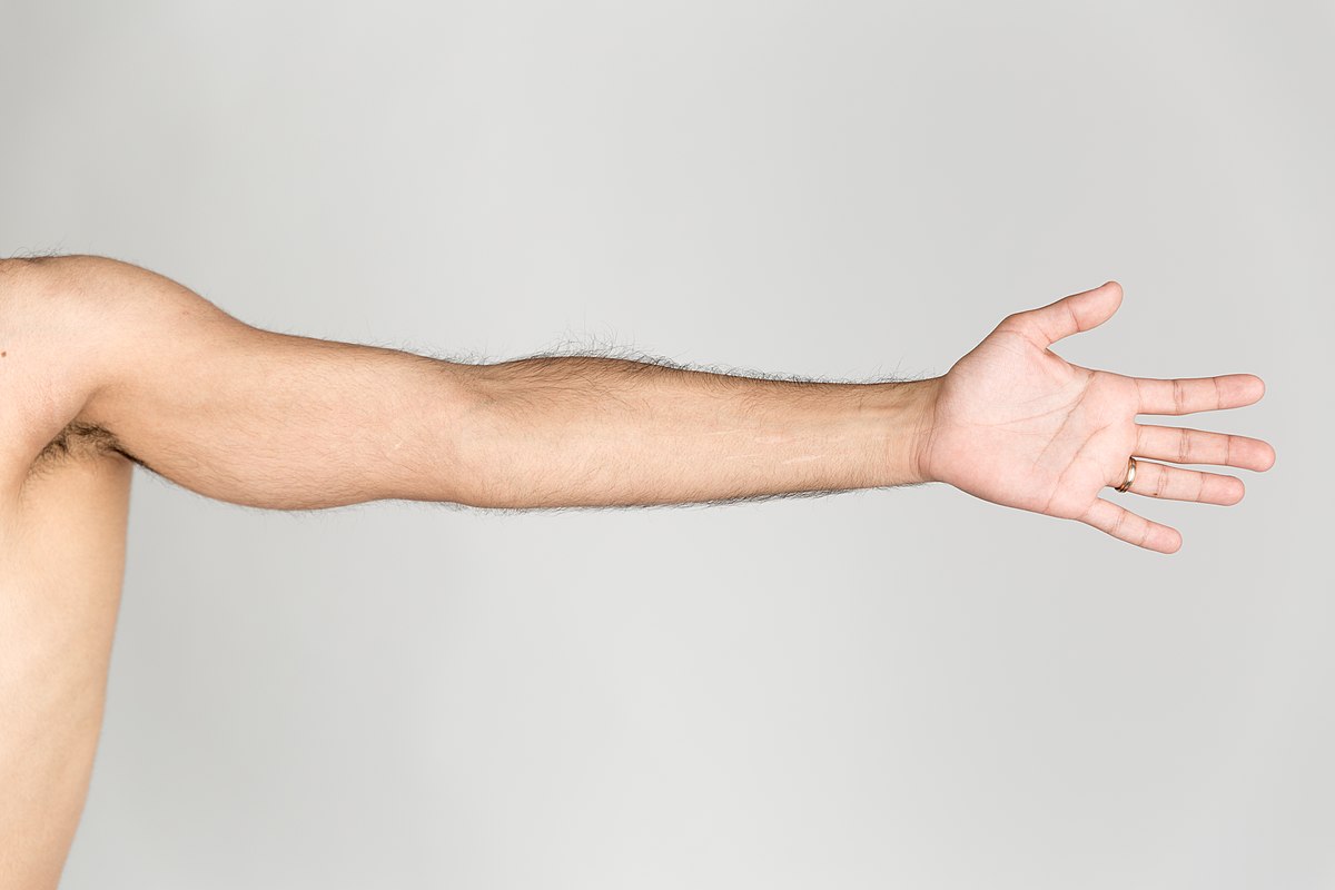
In this article, we will discuss the comprehensive anatomy of the arm. The arm, serving as the connection between the shoulder and the forearm, plays a crucial role in the movements of the hand – the most important limb for human body movement. This section provides a complete description of the components of the arm, from bones to muscles, vessels, and nerves, and finally examines the radiography of the arm.
- Introduction: Comprehensive Anatomy of the Arm
- Anatomy of the Arm Bone
- Dissection of the Arm Tuberosity and Transverse Ligament
- Structure of the Arm Bone Head and Its Joint
- Neck and Shaft of the Arm Bone
- Deltoid Tuberosity and Structure of the Arm Bone
- Inferior Structure of the Arm Bone
- Medial Epicondyle and Trochlea of the Arm Bone
- Joints and Cartilages Associated with the Arm Bone
- Anatomy of the Arm Joints and Bone Depressions Anterior Depressions of the Arm Bone
- Anatomy of the Arm Muscles General Classification of Arm Muscles
- Anatomy of the Arm Nerves
Anatomy of the Arm Bone
The arm bone, or Humerus, is a long bone that articulates at the top with the shoulder blade or Scapula and at the elbow with the forearm bones, namely the upper spoke (Radius) and the lower spoke (Ulna). This bone is the largest bone of the upper limb and is surrounded by the muscles of the arm.
The head of the arm bone (Humeral head) is spherical and is accompanied by two prominences named the Greater tuberosity and the Lesser tuberosity. The Greater tuberosity is located on the outer part where the head connects to the body, and the Lesser tuberosity is present on the anterior or front part of this area.
Dissection of the Arm Tuberosity and Transverse Ligament
Between the small and large tuberosities of the arm, there is a groove called the Bicipital groove. Over this groove, the Transverse humeral ligament exists, creating a tunnel through which the long head of the biceps tendon passes.
Structure of the Arm Bone Head and Its Joint
The head of the arm bone forms about one-third of the surface of a complete sphere and is covered with articular cartilage. This part articulates with the Glenoid fossa of the shoulder blade or Scapula.
Neck and Shaft of the Arm Bone
The area where the head of the arm bone adheres to its body is called the Humeral neck. The middle part of the bone is known as the Humeral shaft, which is cylindrical at the top and becomes broader at the bottom.
Deltoid Tuberosity and Structure of the Arm Bone
In the center of the bone shaft, there is a prominence called the Deltoid tuberosity, which is the attachment site for the deltoid muscle.
Inferior Structure of the Arm Bone
In the lower parts of the arm bone, the cross-section changes from circular to triangular and becomes almost flat and smooth further down. In these areas, prominences called the Lateral condyle and Medial condyle are present.
Medial Epicondyle and Trochlea of the Arm Bone
Attached to the Medial condyle, there is a prominence named the Medial epicondyle. Between the Lateral and Medial condyles, the bone takes on a pulley-like shape (Trochlea).
Joints and Cartilages Associated with the Arm Bone
Anterior, Inferior, and Posterior Surfaces of the Trochlea
The anterior, inferior, and posterior surfaces of the trochlea are covered with articular cartilage and articulate with the upper part of the Ulna bone. The part of the Ulna that articulates with the trochlea is called the Semilunar notch. The anterior and lower surface of the Lateral condyle is more prominent and is covered with the Capitulum, which articulates with the head of the Radius bone.
Anatomy of the Arm Joints and Bone Depressions Anterior Depressions of the Arm Bone
Above the Capitulum and in front of the arm bone, there is a depression called the Radial fossa, which accommodates the head of the Radius bone when the elbow is fully bent.
Above the Trochlea and in front, there is another depression called the Coronoid fossa, which accommodates the Coronoid process when the elbow joint is fully bent.
Posterior Depressions of the Arm Bone
Above the Trochlea and at the back of the arm bone, there is a deep depression called the Olecranon fossa, which accommodates the Olecranon process when the elbow is fully extended.
Anatomy of the Arm Muscles General Classification of Arm Muscles
The muscles of the arm are divided into two groups: the Triceps muscle in the back of the arm, and the three muscles in the front of the arm, which are the Biceps, Brachialis, and Coracobrachialis.
Biceps Brachii Muscle
This muscle is located in the front of the arm and has two heads. The long head starts from the upper edge of the Glenoid cavity and passes through the Bicipital groove. The short head begins from the Coracoid process and extends downwards. The Biceps Brachii muscle is involved in bending the forearm on the arm and in the external rotation of the forearm.
Brachialis Muscle
This muscle is located under the Biceps and adheres to the anterior surface of the arm bone, connecting to the upper and anterior part of the Ulna bone. The Brachialis muscle plays a role in bending the elbow joint.
Coracobrachialis Muscle
This muscle is smaller than the previous two and extends from the Coracoid process downwards, adhering to the medial edge of the arm bone shaft. The function of this muscle is to bring the arm forward and closer to the trunk.
Anatomy of the Triceps Brachii Muscle
The Triceps Brachii muscle is located at the back of the arm and has three tendinous heads. The long head starts from below the Glenoid cavity, and the lateral and medial heads adhere to the upper and posterior surface of the arm bone. This muscle connects at the bottom to the Olecranon process of the Ulna bone and plays a role in straightening and extending the elbow joint. Additionally, it can move the arm backward and bring it closer to the trunk.
Anatomy of the Arm Nerves
Musculocutaneous Nerve
This nerve branches off from the brachial plexus and controls the activity of three muscles in the arm: the Biceps, Brachialis, and Coracobrachialis. In the lower parts of the arm, it provides sensation to a portion of the skin of the forearm.
Radial Nerve
The Radial nerve branches off from the posterior cord of the brachial plexus and activates the Triceps muscle in the arm. This nerve is located inside the upper parts of the arm and moves towards the back of the arm bone. Below the elbow, it controls the movement of some forearm muscles and the sensation of a part of the hand.
Median and Ulnar Nerves
These two nerves, after emerging from the brachial plexus, move downwards on the inner side of the arm. They do not perform any specific function in the arm but reach the forearm and hand, where they control sensation and movement.

To make an appointment or get an online consultation with Dr. Nader Motallebi Zadeh, Limb lengthening surgeon, proceed here.
