Overview of Humerus Fracture
A fracture of the humerus (upper arm bone) is one of the common types of fractures, accounting for about five percent (one-twentieth) of all body fractures. This type of fracture is less common in children compared to forearm fractures.

- Overview of Humerus Fracture
- Mechanism and Causes of Fracture
- Symptoms of Humerus Fracture
- Nerve Damage Associated with Fracture
- Diagnostic Methods for Humerus Fracture
- Initial Care for Humerus Fracture
- Immediate Actions at the Accident Site
- Non-Surgical Treatments for Arm Fractures
- Non-Surgical Treatment Options
- Use of Casting in Treating Humerus Fractures
- Mechanism and Purpose of Casting
- Care Instructions for Patients
- Next Steps in Treatment
- Use of Splints or Braces Instead of Casts
- Post-Casting Actions in Arm Fracture Treatment
- Follow-up Treatment and Possibility of Fragment Displacement
- Necessary Care During Treatment
- Special Considerations in Setting Broken Fragments
- Surgical Treatment of Humerus Fractures
Mechanism and Causes of Fracture
The primary mechanism of a humerus fracture is falling on an outstretched hand. During a fall, a person instinctively extends their hand forward in a stretched position. The impact of the hand hitting the ground transfers force to the arm, leading to a fracture. Additionally, car accidents and falls from heights are other causes of humerus fractures. These fractures usually occur in the middle third of the humerus bone.
Symptoms of Humerus Fracture
The symptoms of a humerus fracture are usually obvious and identifiable. At the moment of impact and fracture, the patient may hear or feel the fracture and immediately experience severe pain in the arm. The arm becomes deformed and swollen, and the skin over the fracture site gradually turns bruise-colored. The patient also feels severe pain at the fracture site, which intensifies with arm movement. Often, patients try to support the injured arm and forearm with their healthy hands.
Nerve Damage Associated with Fracture
In some cases of humerus fractures, due to the proximity of the radial nerve to the bone, paralysis of this nerve can occur. Paralysis of the radial nerve can manifest as a loss of sensation in the back of the hand and an inability to lift the wrist and fingers.
Diagnostic Methods for Humerus Fracture
In humerus fractures, especially in the lower parts, there is a possibility of damage to the radial nerve. During the examination, the doctor carefully assesses the function of this nerve. The patient is evaluated for the ability to move fingers and the wrist, as well as for the sense of touch on the back of the hand.
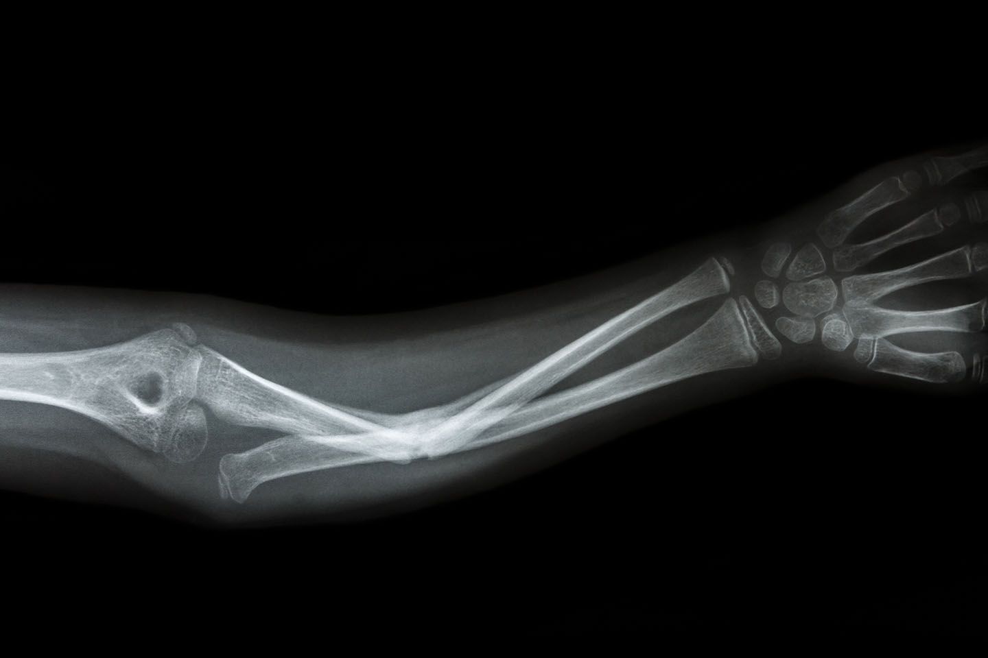
For an accurate diagnosis of the fracture and its type, radiography is used. Simple radiography is primarily used to confirm a humerus fracture. Additionally, the doctor may take images of the shoulder and elbow areas to ensure there is no damage to these joints.
Initial Care for Humerus Fracture
When encountering someone with a humerus fracture, first ensure that they are away from the accident scene and not in further danger. If severe bleeding or the possibility of other fractures in the body is observed, immediately contact emergency services. If the broken bone is protruding from the wound, do not attempt to push it back in.
Immediate Actions at the Accident Site
Ensure that the patient’s breathing and pulse are normal. If there is swelling or bleeding, reduce it by gently applying pressure and elevating the injured limb above heart level. Also, cover any potential wounds with a clean cloth or gauze to prevent further contamination. Immobilize the broken arm by tying it with a cloth around the neck and keeping it still relative to the torso. Then, transport the injured person to the nearest medical facility.
Non-Surgical Treatments for Arm Fractures
Non-Surgical Treatment Options
In many cases, arm fractures can be treated without the need for surgery. These treatments usually involve the use of specific casts such as a “hanging cast,” a U-Slab plaster splint, or arm splints. Casting is the most common non-surgical treatment method for arm fractures.
Use of Casting in Treating Humerus Fractures
Casting is a common method for treating arm fractures. In this method, the patient’s arm and forearm up to the palm are covered with a cast. The patient’s elbow joint is positioned at a 90-degree angle, while the wrist joint remains straight without deviation. The cast around the forearm is suspended from the patient’s neck with a cloth strap.
Mechanism and Purpose of Casting
The main purpose of this cast is to separate the broken arm fragments and allow the fracture to be set by the weight of the cast itself. This process works best when the patient is standing or sitting, as the weight of the cast causes the broken fragments to separate and set correctly.
Care Instructions for Patients
During the first two weeks of treatment, it is recommended that the patient remains standing or sitting and even sleeps in a semi-sitting position. The patient should also avoid removing the cast from their neck. After three weeks, the broken fragments adhere to each other and no longer move relative to each other.
Next Steps in Treatment
After about a month, the treating physician removes the cast and applies a special plastic brace around the arm. The use of this brace allows the patient to move their wrist and elbow. Complete fracture healing typically occurs within one and a half to two months. After this period, the patient should continue necessary precautions to prevent re-injury to the arm.
Use of Splints or Braces Instead of Casts
In some cases, instead of using a hanging cast, a plaster splint or brace is used for the initial treatment of the arm fracture.
Post-Casting Actions in Arm Fracture Treatment
If non-surgical treatment is chosen, the doctor will take weekly X-rays of your arm during the first few weeks of treatment. This is due to the possibility of the broken fragments shifting after the start of treatment. If the displacement is within acceptable limits, non-surgical treatment continues; otherwise, the doctor may recommend surgery.
Follow-up Treatment and Possibility of Fragment Displacement
There is a possibility of secondary displacement of the broken fragments up to three weeks after the fracture. Therefore, the doctor takes weekly X-rays of the fracture site for three weeks. If the broken fragments do not shift during this period, the likelihood of future displacement is very low.
Necessary Care During Treatment
Healing of an arm fracture may take several weeks to months. During this time, repeated contraction of the arm muscles is very important and should be done under the supervision of a doctor or physiotherapist. Also, gradual movements of the shoulder and elbow should be started to prevent joint stiffness. The timing and manner of these movements are determined by the doctor.
Special Considerations in Setting Broken Fragments
In closed reduction, the fracture fragments usually do not align precisely, and this is not necessary. Even if subsequent radiographs show that the fragments are not in their exact place, there is no need to worry, and you should trust your treating physician’s assessment.
An arm fracture, even with some displacement, heals correctly and does not affect the arm’s appearance or function. Sometimes, accepting some displacement is better than undergoing surgery. However, it should be noted that not all degrees of displacement are acceptable, and surgery is necessary in cases where the displacement is more than usual.
Surgical Treatment of Humerus Fractures
Indications for Surgical Treatment
In most cases, fractures of the humerus can be treated without surgery, using only casting or bracing. However, sometimes a doctor may decide to use surgery. The main reasons for this decision include:
- Failure of closed reduction and the inability of non-surgical treatment to heal the fracture properly.
- Presence of fractures alongside other physical injuries or fractures in other limbs.
- Fractures are accompanied by nerve or vascular problems.
- Open fractures.
Surgical Treatment Methods
Surgical treatment usually involves open reduction of the fracture and stabilizing the broken pieces with screws and plates. After surgery, patients can typically move their shoulder and elbow from the next day. Performing these movements a few days after surgery is essential to prevent limited range of motion in the shoulder and elbow joints. Until the doctor permits, the patient should not lift anything with the injured limb, but should perform joint movements.
Lifting Ability Post-Surgery
Usually, a few months after surgery, the fracture site becomes strong enough for the patient to lift objects with the injured limb. Undertaking heavier activities generally requires at least six months.
Post-Healing Care
After the fracture has healed, it is usually not necessary to remove the screws or plates from the arm, except in specific cases determined by the doctor. In some cases, treatment may involve the use of an intramedullary nail. In this method, closed reduction is performed, and a long metal rod is inserted through a small incision in the shoulder or elbow area into the medullary canal of the humerus, which may be attached to the humerus with a few screws.
Use of External Fixators
Sometimes, external fixators are used to treat fractures of the humerus. This device is particularly used in treating open fractures of the humerus or fractures accompanied by infection.
Complications of Humerus Fractures
Humerus fractures usually heal without specific problems, but sometimes potential complications exist. The most important of these complications include:
- Nerve Damage: Damage to the radial nerve occurs in about ten percent of arm fractures. The radial nerve, close to the middle and distal parts of the humerus, can easily be damaged during a fracture. The main sign of radial nerve damage is paralysis of the muscles that lift the wrist and fingers and reduced touch sensation in the skin on the back of the hand.
- Treatment of Radial Nerve Damage: The initial treatment for radial nerve damage after a humerus fracture is observation and care, as many of these injuries are neuropraxia and heal on their own. If the doctor does not observe improvement after a specific period, surgery is performed. In surgery, the damaged nerve site is exposed and repaired if necessary.
- Tendon Transfer in Case of Unsuccessful Nerve Repair: If nerve repair is not possible, another treatment method is tendon transfer. In this method, the surgeon changes the attachment of some tendons in the upper limb to enable movements that the hand is incapable of performing.
- Non-Union of Bone Fracture: Sometimes, a bone fracture does not heal, even after sufficient time. This problem can occur in both non-surgical and surgical treatments. Treatment of non-union of a humerus fracture usually involves surgery and bone grafting. In this method, the fracture site is usually fixed using a plate, or if a plate has been used previously and the fracture site is moving, the plate is replaced.
- Malunion of Bone Fracture: In non-surgical treatments of humerus fractures, the broken pieces usually do not fuse together precisely, but this situation usually does not create a significant change in the appearance or function of the arm. Even a few centimeters of shortening of the arm usually does not cause a problem in the function of the upper limb.
- Bone Infection: Bone infection is more common in open fractures of the humerus and is more likely in patients who have undergone surgery. Symptoms of infection usually include redness and swelling of the arm and discharge of pus from the fracture site. The patient may also have general symptoms such as fever. Treatment of this complication usually involves the use of injectable antibiotics and surgery to remove dead tissues and replace or remove the plate or intramedullary nail.

To make an appointment or get an online consultation with Dr. Nader Motallebi Zadeh, Limb lengthening surgeon, proceed here.


