Osteotomy
A bone-cutting technique called an osteotomy is used to straighten and restructure your bones and joints. Common surgery locations include your jaw, elbow, spine, shoulder, hips, knees, legs, and toes. There are numerous surgical methods and method modifications. In order to get you up and moving again with less pain and discomfort, your surgeon will explain the best treatment choice for your particular bone issue.
What is an osteotomy?
In order to restructure or realign your bones, a surgical technique called an osteotomy involves cutting bone (and occasionally adding bone tissue).
Any bone on your body, including the ones in your jaw, elbow, spine, shoulder, hips, knees, legs, toes, and feet, can undergo an osteotomy with the help of your surgeon.
It is a remedy for conditions that affect your joints, which are where two bones meet.
Many osteotomy technique subtypes and variants exist. They are frequently given names based on the surgeon who developed the technique or the way the bone is cut, reshaped, or modified.
Why it is performed?
An osteotomy is carried out by your doctor to:
- Adjust your bones’ angle, bow, or rotation.
- Align a joint that is misaligned or malformed.
- bone lengthening or shortening.
- mending a broken joint
- Move your weight away from a joint’s damaged portion and toward a region with more normal or healthy cartilage.
- reduce arthritic pain (especially in the knee and hip).
- Avoid having joint replacement surgery when you’re young and active.
- Restore or resolve other specific bone problems.
- Limb lengthening surgery
How do I prepare for an osteotomy?
To assess your general health, your surgeon could request several standard tests. They may consist of:
- Blood tests to determine the levels of specific blood components and the efficiency of your organs.
- Urine tests to evaluate your overall health and spot conditions like infection or diabetes that prevent bone regeneration.
- Electrocardiogram to examine the electrical activity of your heart.
- Before surgery, get a chest X-ray to check sure your lungs are healthy.
- To establish the surgical plan, X-rays or computed tomography (CT) scans are used to visualize your bones and joints. The precise size, dimensions, and angle of the bone portion to be removed must be determined by your surgeon. On occasion, a 3D model of the surgery is created on a computer.
What happens during an osteotomy operation typically?
You will first experience anesthesia. Your doctor might decide to:
- Just numb the surgery site (with regional anesthesia).
- From your waist down, numb your body (with spinal anesthesia).
- You’re put to sleep (with general anesthesia).
- only numb the surgical location (with local anesthesia).
The region surrounding the surgical site is then sterilised by your surgical team using an antibiotic solution. A surgical drape covers the area.
A skin incision is made by your surgeon. To delineate the area of bone to be removed, they employ guide wires (often wedge-shaped, but depends on the procedure). The specified bone section is removed using a specialized surgical saw.
The gap in your bone is then filled by bringing the borders of the bone together after your surgeon removes the affected or diseased portion. Depending on the details of your individual osteotomy treatment, a bone graft may occasionally be put into the area where the bone was removed. Once the bone heals, it may be realigned using pins, screws, plates, staples, or rods. This metal may be positioned either temporarily or permanently.
What various osteotomies are there?
Numerous different kinds of bone and joint issues can be resolved with an osteotomy. They consist of:
Jaw osteotomy
The lower jaw (mandible) or upper jaw (maxilla) bones are realigned with the remainder of the head and/or teeth during a jaw osteotomy. An open bite, difficulty chewing or swallowing, excessive tooth wear, a receding chin, an overbite, or an underbite can all be fixed by jaw osteo’tomy. To realign your teeth with your jaw before or after surgery, you might need to wear braces.
The following are terms or classifications of jaw osteotomies:
- osteotomy of the mandible. This procedure involves your lower jaw.
- osteotomy of the mouth. This procedure involves your upper jaw.
- osteotomy LeFort. Your upper jaw is involved in a midface fracture that is treated using this specific surgical approach.
- osteotomy with a sagittal split. This is a particular kind of jaw surgery to realign your lower jaw with your teeth.
Chin osteotomy
Your chin is reshaped through a chin osteotomy. Often, your surgeon will perform a chin osteo’tomy to lengthen your chin, shorten a narrow chin, or fix a vertically short chin. In certain circumstances, this procedure is an alternative to getting a chin implant because it moves your chin forward or in other ways.
Chin osteotomies entail cutting and repositioning your jaw bone. Your mouth is used as the site of the incision by the surgeon. During a few months or longer, your lips can lose feeling.
Elbow osteotomy
An elbow osteotomy corrects abnormalities with the elbow joint that affect how your lower arm is aligned. Your arm is either tilted too far away from your torso or hangs too low (cubitus varus) (cubitus valgus). These unnatural poses alter the elbow’s carrying angle. Several osteo’tomy procedures exist, depending on the precise issue.
Spinal osteotomy
Your spine’s curvature are aligned with the help of a spinal osteotomy. The inherent curves of your spine assist in placing your body’s center of gravity over your pelvis. The lower portion of your torso’s bone structure is called your pelvis. Your spine becomes misaligned if one or more spinal segments have an excessive amount of curvature or insufficient amount of curvature.
Internal organs may be under pressure, you may feel weary, and you may experience discomfort. A spinal osteotomy aims to restore balance, ease discomfort, and stop the deformity from returning or getting worse.
To fix an angle issue, such as a persistent forward bend that causes your chin to rest on your chest, your surgeon may try osteo’tomy. In ankylosing spondylitis, this occurs. In extreme circumstances, this form of arthritis can cause your spine’s discs to fuse together.
The most common spinal osteotomy techniques include:
- Osteotomy of the posterior column This procedure corrects a forward-curving upper spine and an inwardly arched lower back (lordosis) (kyphosis). Ponte osteotomy is a particular method for treating kyphosis.
- Osteotomy by Smith-Petersen. This particular method corrects lordosis. Your spine would slant further toward the back if a piece of bone were removed from the back.
- osteoporotic pedicle subtraction. During this procedure, your lower spine’s more severe (more arching) lordosis is corrected.
- osteotomy of the bone-disk-bone. Your spine is corrected above and below a disk with this procedure. Surgery is performed to remove the disk and its nearby endplate (s).
- spinal column removal. This procedure, which offers the greatest correction, involves the total removal of one or more vertebrae. Your surgeon will use a graft or metal cage to fuse this part of your spine because so much bone was removed.
Hip osteotomy
Your hip socket (acetabulum) and/or thighbone’s head are reshaped during a hip osteotomy (femur head). Your hip joint is made up of a ball and socket. To realign the weight-bearing surfaces of the joint, your surgeon makes cuts, forms, or partial removals of bone tissue.
The following are the primary kinds of hip osteotomies:
- osteotomy around the acetabulum. This osteo’tomy is carried out by your surgeon to treat hip dysplasia, which is a condition in which the ball portion of your upper thigh bone is not completely covered by the hip socket. Your hip bone is sliced through during surgery, repositioned, and screwed into place.
- Osteotomy of the femur In order to restore hip function, this procedure entails cutting and straightening the femur, which is the upper thigh bone.
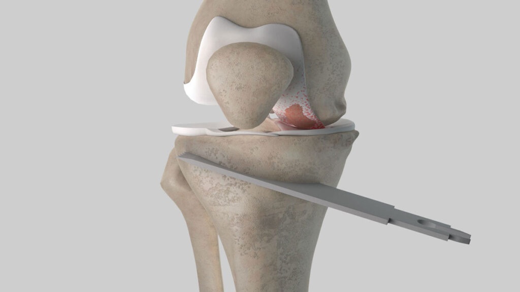
Knee osteotomy
The shinbone (tibia) or thigh bone, which meet under the kneecap, are cut and reshaped during a knee osteotomy (femur). Usually, it’s done to repair arthritis damage. The procedure realigns your knee joint, transferring pressure and weight from the injured side of your knee to the healthy side. Osteotomy of the knee is typically performed when osteoarthritis is first diagnosed and just one side of the knee joint has been damaged.
The site of the knee osteotomy is determined by the injury. For instance, cartilage injury usually occurs on the inside of your knee following a high tibial osteo’tomy. In order to straighten your leg and stop the growth of arthritis, the procedure entails either removing a wedge of bone from the outside of your knee or opening up a wedge of bone on the inside of your knee.
Knee osteotomies come in several forms, such as:
- osteo’tomy of the tibia With this procedure, you can rectify your bowlegged posture, which puts too much strain on the inside of your knee.
- osteo’tomy of the high tibia. Patients with knee arthritis who undergo this operation have their knee joint realigned. The necessity for a partial or full knee replacement may be avoided or postponed with this procedure.
- Osteo’tomy by Fulkerson. This particular method entails removing a portion of your tibial tubercle (a specific area on your tibia). This alters the site of the kneecap tendon’s attachment to the tibia, relieving strain on the joint and preventing dislocation.
Big toe and foot osteotomies
Your big toe can be straightened by having a big toe (hallux) osteotomy. An operation to restructure your foot to correct flat feet or a higher-than-normal arch is called a heel (calcaneus) osteotomy.
Many of the particular osteotomy methods used to address foot issues include:
- Osteotomies at Akin and Chevron. These treatments fix bunions-related mild to moderate big toe alignment issues (hallux valgus).
- Osteotomy Dwyer. Your foot is reshaped during this treatment to lower an excessively high arch.
- osteotomy by Weil. This technique eliminates pain under the ball of your foot and claw toe.
- osteotomy of cotton. Your foot will develop an arch as a result of this surgery.
What dangers come with having an osteotomy?
Among the dangers of an osteotomy are:
- difficulties with anesthesia
- Infection.
- clots of blood.
- artery or nerve damage.
- bones that don’t correctly heal or align as they do.
- stiffness and joint inflammatory disease.
- chronic discomfort
- skin scars.
What may I anticipate when I recover?
The type of osteotomy, the precise surgical technique, the extent and seriousness of the bone injury, as well as your stamina and determination, all affect how well you recover.
Your bone needs time to mend. You’ll have pain where your procedure was performed.
You could require a cast, splint, or crutches to restrict bone and joint movement, keep weight off the operated bone, and promote proper bone healing. For a few weeks to a few months, you will likely need to use crutches or wear a cast or splint.
Even if you are wearing a cast or splint, physical therapy will start immediately following your operation. Your joint strength and range of motion are improved by physical therapy.
If you had surgery on your knee or hip, you might need to wear crutches for a few months. Physical therapy will be used to help you restore your strength and balance.
Your jaw is wired shut after a jaw “osteotomy”, and you must follow a liquid diet for six weeks. During two to six weeks after an osteotomy on your big toe, you cannot drive or wear shoes.
What can I do to promote healing?
You can facilitate healing by:
- not a smoker. Nicotine slows down the healing process and could hinder proper bone fusion.
- consuming a balanced, largely plant-based diet, such as the Mediterranean diet.
- observing the advice of your healthcare expert.
- the upkeep of a healthy weight.
What results can be anticipated?
Your results are dependent on your overall health, the seriousness of your bone condition, the particular treatment you had, and the skill of your surgeon. Your surgeon will go over the particulars of your procedure and what to anticipate. Do not be afraid to express any worries or ask questions to your surgeon.