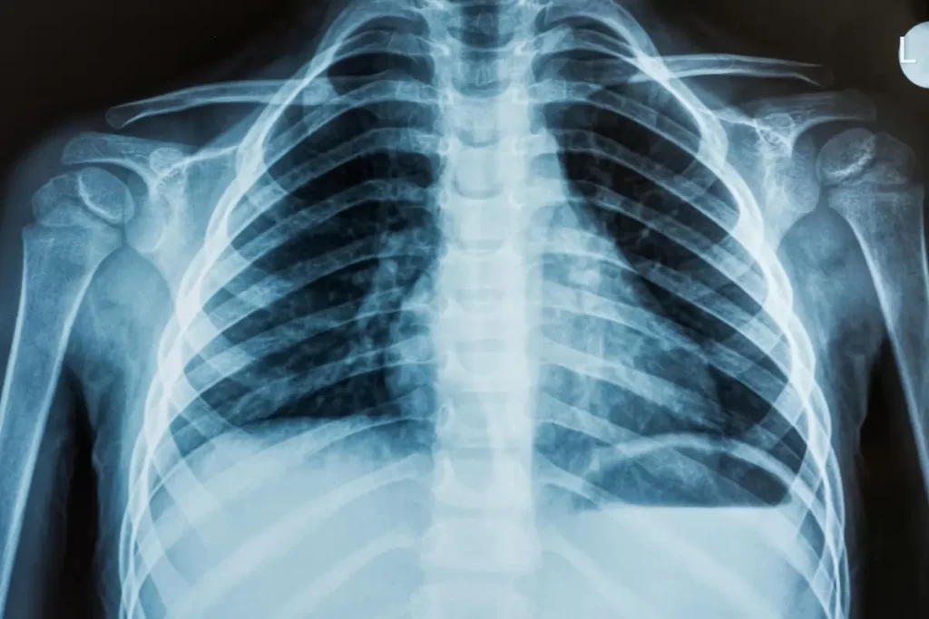
X-Ray
An X-ray is a type of imaging test that captures images of the soft tissues and bones. Safe radiation doses are used by X-rays to produce these images. The photos aid in the diagnosis of numerous illnesses and the formulation of treatment plans. X-rays are typically used by medical professionals to assess fractured bones, dislocated joints, and other bone ailments.
What is an X-ray study?
An X-ray scan, often known as a radiograph, is a type of radiology that takes images of your organs and soft tissues like your bones. These images are produced by X.rays using low levels of energy. Your doctor can detect diseases and devise treatment plans with the aid of the photographs.
Providers typically utilize X-rays to check for fractures (broken bones). Yet Xray scans can aid medical professionals in the diagnosis of a variety of wounds, conditions, and illnesses. Xrays are a secure and reliable method for medical professionals to assess your health.
Who may require an X-ray?
You can have an X-ray if you’re any age, even a baby. Inform your provider if you think you could be pregnant before having an X-ray. Your fetus could be harmed by Xray radiation.
Your doctor might ask for an X.ray to:
- Inspect for shattered bones (fracture).
- Determine the origin of symptoms like pain and edema.
- Check your body for foreign items.
- Check your bones, joints, and soft tissues for structural concerns.
- Create and assess treatment plans.
- Provide regular cancer and other disease screenings.
- Check the recovery from limb lengthening surgery
What are the types of X-ray studies?
Several X-ray procedures use photography to capture various internal body regions. To improve the clarity of the images, certain X-rays employ contrast material, commonly known as dye. Among the most popular X.ray procedures are:
- An abdominal X.ray provides images of your bladder, stomach, liver, and kidneys. It aids medical professionals in the diagnosis of ailments including bladder and kidney stones. Certain specialized abdominal X.rays, like a barium enema, use specific dyes (referred to as contrast) to assess various components of the digestive system.
- Bone X-ray: Your doctor will use a bone X.ray examination to check for arthritis, displaced joints, and shattered bones (fractures). Bone cancer or infections can also be visible in the images from bone X.rays. An X.ray of the spine examines its tissues and bones.
- Chest X-ray: This examination searches for pneumonia-like anomalies in the heart, lungs, and chest bones.
- Frequent dental X-rays provide your doctor the opportunity to examine your teeth and gums, search for infections, and look for cavities.
- Organs and soft tissues are depicted in moving images during a fluoroscopy (such as your intestines). Your organs are seen moving on a screen by your doctor (kind of like an X-ray movie). Fluoroscopy is frequently used in GI X.ray tests.
- CT scan: A radiological procedure that produces cross-sectional images of bones, organs, and tissues using X.rays and a computer. You slide through this donut-shaped contraption as it takes pictures.
- Mammogram: Healthcare professionals use mammograms to examine breast lumps, capture X.ray images of breast tissue, and identify breast cancer.
What is an X-ray with contrast material?
Contrast media is used with some X.rays (also called contrast agent or dye). The contrast substance is offered as a liquid, powder, or tablet. Before the X-ray, your doctor gives you the contrast material. You might get the contrast material, depending on the kind of X.ray:
- Orally (by mouth) (by mouth).
- through a shot delivered intravenously (IV), for example.
- By placing it inside of your rectus (enema).
You can get a brief flush or warm feeling after receiving the dye through an IV injection from your healthcare professional. A metallic taste can sometimes be detected in the mouth. These side effects disappear quickly.
To enable your provider to view soft tissues and other structures more clearly on an X.ray study, a contrast agent is used.
How does an X-ray study work?
Your body is exposed to radiation rays during an X.ray. You can’t feel or see radiation beams, and they are invisible. Your body is exposed to the beams, which then produce an image on a nearby X.ray detector.
Your body’s bones, soft tissues, and other structures all absorb radiation differently as the beams pass through it. Bones and other solid or dense materials readily absorb radiation, giving them a dazzling white appearance on the image. Soft tissues, such as organs, appear in various colors of gray on X.rays because they don’t absorb radiation as well.
How do I prepare for an X-ray?
Inform your doctor about your medical history, allergies, and current medication intake. Inform your physician if you are breast-feeding, suspect you may be pregnant, or are pregnant before receiving an X.ray.
With a bone X-ray, you often don’t need to do anything in advance. Your healthcare provider can require you to:
- Avoid using creams, lotions, or perfume.
- Take off any jewelry, hairpins, or hearing aids that are made of metal.
- Put food and liquids away several hours in advance (for GI X.rays).
- Before the X.ray, dress comfortably or change into a gown.
What should I expect during an X.ray?
Your doctor may ask you to stand, sit, or lie down on a table depending on the type of X.ray that needs to be taken.
Your healthcare professional can advise you to hold motionless while moving your body or limbs around during the X-ray. To prevent hazy visuals, you might need to hold your breath for a short period of time.
Children occasionally struggle to remain motionless for long enough to make clear photographs. A restraint may be advised by your child’s doctor during an X-ray. The immobilizer or strap keeps your infant immobile and lessens the number of retakes that are necessary. The restraints are safe for your youngster and do not damage them.
What should I expect after an X-ray?
Drink a lot of water to help your body rid itself of the contrast material if you received it before your X.ray. Contrast dye may cause some persons to have the following adverse effects:
- sickness or vomiting
- diarrhea or cramps in the stomach
- Headaches.
- Occasionally, contrast material allergic reactions can happen. An allergic reaction to contrast dye is more prevalent in people with allergies or asthma. If you have any unusual symptoms, call your provider right away and discuss your risk of having a reaction with them.
What are the risks of an X.ray?
Despite the fact that X.rays involve radiation, which can lead to cancer and other health issues, there is a negligible danger of radiation overexposure during an X.ray. Different X.rays employ different radiation dosages. In general, X.rays are efficient and safe for patients of all ages.
Your fetus could be harmed by X.ray radiation. Your doctor might decide on another imaging test if you’re pregnant, such an MRI or ultrasound.
When should I be made aware of my X.ray results?
An X-ray of a bone typically yields results right away. After the X.ray, your doctor might discuss your results with you. It could take longer to get the results from other X.ray tests (such a GI test). Find out from your provider when you can anticipate results.
When should I call my doctor?
Contrast materials seldom cause allergic responses. Up to a day or two following the X-ray, symptoms may start to show. Call your provider if any of the following occur after receiving contrast material before your X.ray:
- Itching, hives, or skin rash.
- Headaches.
- sickness or vomiting
- Shortness of breath or difficulty breathing.