Overview: Calcific tendonitis, characterized by calcium deposits in the shoulder tendons leading to inflammation and pain, is often observed in individuals over the age of 40.
Functional Anatomy of Calcific Tendonitis
Anatomical Details: In calcific tendonitis, calcium deposits in the tendons of the rotator cuff. This group consists of four muscles that attach to the humerus and hold it firmly in the glenoid cavity. Primarily, calcium deposition occurs in the tendon of the supraspinatus muscle.
Mechanisms of Calcific Tendonitis Development
Causes: This condition arises from two mechanisms:
- Reduced Blood Flow with Aging: As age increases, blood flow in the rotator cuff decreases, weakening the tendon. This weakness leads to microscopic tears in the tendon, which become more extensive over time. The body, in an attempt to repair these tears, causes calcium to deposit on the tendon.
- Inflammation of the Rotator Cuff Tendon: For unknown reasons, the rotator cuff tendon becomes inflamed, and this inflammation leads to calcium deposition. Over time, the body absorbs these calcium deposits, a process that causes severe pain in the shoulder area. Gradually, the inflammation of the tendon decreases, and the condition improves.
Unknown Cause of Calcium Deposition
Uncertainty of Cause: The exact reason for calcium deposition in these conditions remains unknown.
Symptoms of Calcific Tendonitis
Symptomatology: Symptoms of this condition include shoulder pain, which usually begins during the body’s reabsorption of calcium. Initially, the pain may be mild or even non-existent. This pain may intensify with shoulder movement, especially when raising the hand towards the head. Sometimes, the pain becomes so severe that it disrupts the person’s sleep.
Diagnostic Methods for Calcific Tendonitis
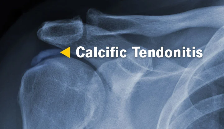
Diagnosis: This condition is typically diagnosed using simple radiography. In this method, calcium deposits appear as white spots on the top of the humerus.
Treatment of Calcific Tendonitis
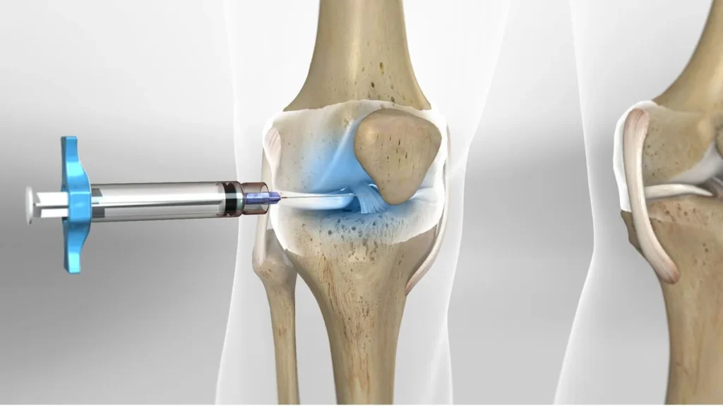
Initial Treatment: The primary treatment includes non-surgical methods such as reducing physical activities and resting the shoulder. During this period, the use of anti-inflammatory drugs like piroxicam or ibuprofen can help reduce pain. Another treatment method involves the orthopedic doctor removing the calcium using a needle and injecting saline into the site of calcium accumulation.
Physiotherapy: Physiotherapy, including the use of heat or cold, ultrasound, and shock wave therapy, can also help reduce pain. These methods can break down the deposited calcium into very fine pieces, allowing better absorption by the body.
Surgical Intervention: If the aforementioned treatments are ineffective, surgery is considered. This surgery is usually performed arthroscopically, during which the deposited calcium is removed from the tendon.

To make an appointment or get an online consultation with Dr. Nader Motallebi Zadeh, Limb lengthening surgeon, proceed here.