Introduction to the Elbow Joint
The elbow joint is a complex hinge joint composed of multiple components. In this article, we will examine in detail the bones, surrounding tissues, as well as the vessels and nerves associated with it. The final section will provide information related to elbow radiography.
Anatomy of the Bones of the Elbow Joint
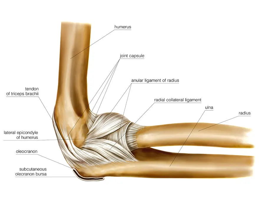
The elbow joint is where the three main bones meet. At the top of the joint is the humerus, and below are the two forearm bones named the radius and ulna. The radius bone is located on the outer side of the elbow, near the thumb, and the ulna on the inner side, near the little finger.
The lower end of the humerus in the elbow area becomes bulky and prominent. This prominence can be divided into two major parts: the lateral condyle on the outer part and the medial condyle on the inner part. Near the medial condyle, there is another prominence called the medial epicondyle.
Changes in Bone Shape Between the Lateral and Medial Condyles
Between the lateral and medial condyles, the shape of the bone changes and forms a pulley-like structure called the trochlea. The anterior, inferior, and posterior surfaces of the trochlea are covered with articular cartilage and articulate with the upper part of the ulna.
Semilunar Notch and Capitulum
The part of the ulna articulating with the trochlea is known as the semilunar notch. The front and lower surface of the lateral condyle is slightly more prominent and covered with cartilage. This part, called the capitulum, articulates with the head of the radius.
Radial and Coronoid Fossae
Above the capitulum and at the front, there is a depression known as the radial fossa or radial fossa, which accommodates the head of the radius when the elbow is fully bent. Also, above the capitulum and at the front, there is another depression called the coronoid fossa, where the coronoid process fits when the elbow is fully bent.
Olecranon Fossa
Above the trochlea and at the back, there is a deep depression called the olecranon fossa. When the elbow is fully extended, the olecranon process fits into this depression.
Semilunar Notch and Humeroulnar Joint
The upper part of the ulna is shaped like the letter F, creating a semicircular bone at the top of the bone. This semicircle is called the semilunar or trochlear notch. The semilunar notch, located next to the trochlea of the humerus, forms a hinge joint called the humeroulnar joint. The inner or concave surface of this semicircle is covered with articular cartilage.
This semicircle has two upper and lower edges; the larger upper edge is called the olecranon, and the lower edge is called the coronoid process.
Structure of the Head of the Radius
The upper side of the radius has a prominence known as the head of the radius. The part that connects the head of the radius to its body is called the neck of the radius.
Structure of the Head of the Radius Bone
Head of the Radius Bone
The head of the radius bone is shaped like a circular disc and is covered with articular cartilage. The upper surface of this disc is concave.
Humeroradial Joint
The concave surface of the head of the radius bone meets the convex surface of the capitulum of the humerus, forming the humeroradial joint.
Proximal Radioulnar Joint
Next to the outer edge of the semilunar notch, there is a concave depression that sits alongside the disc-shaped head of the radius bone, forming the proximal radioulnar joint.
Anatomy of the Elbow Joints
Humeroulnar Joint (Humeroulnar)
This joint is located between the humerus and ulna. As the most significant and largest part of the elbow, it is a hinge joint that allows for bending and straightening movements of the elbow. The humeroulnar joint is located on the inner side of the elbow, where the little finger is positioned.
Humeroradial Joint (Humeroradial)
This joint is between the humerus and radius and is situated on the outer side of the elbow, where the thumb is located. The humeroradial joint facilitates both hinge movements (bending and extending of the elbow) and pivot movements (rotation of the radius around its longitudinal axis).
Supination and Pronation Movements
The rotation of the radius bone enables movements of supination (palm facing upwards while the elbow is bent at 90 degrees) and pronation (back of the hand facing upwards in the same position).
Proximal Radioulnar Joint
This joint is between the radius and ulna bones at the upper part. The radius and ulna, which are the two long bones in the forearm, articulate with each other in three areas (upper, middle, and lower). The proximal radioulnar joint also plays a role in the supination and pronation movements of the forearm.
Articular Cartilage of the Elbow Joint
The articular surfaces of the capitulum, trochlea, the inner surface of the semilunar notch, and the upper surface of the head of the radius bone are covered with hyaline cartilage. This cartilage is a smooth, slippery, and white layer that facilitates movement in the joint. Articular cartilage, by creating a smooth and slippery surface, enables the easy movement of the bones that form the joint over each other.
Elbow Joint Capsule
The elbow joint capsule is a thick and sturdy tissue membrane that surrounds the joint. This capsule resembles a cylindrical barrel without the top and bottom bases, with both ends open.
The upper circle of the capsule wraps around the lowermost part of the humerus, and the lower circle adheres around the necks of the radius and ulna bones. This structure creates a completely enclosed space where the lower end of the humerus and the upper ends of the radius and ulna bones are located.
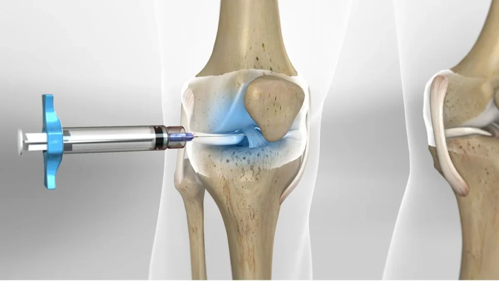
Synovial Membrane of the Elbow Joint
The synovial membrane is a thin layer that covers the inner surface of the joint capsule. Its primary function is the secretion of synovial fluid.
Synovial Fluid of the Elbow
Synovial fluid is a clear, thick, and viscous fluid similar to egg white, secreted by the synovial membrane. This fluid has two main functions:
- Synovial fluid makes the surface over the articular cartilage sticky and slippery, similar to oiling in machines, which aids in smoother joint movement.
- Synovial fluid is rich in nutrients and oxygen, which helps nourish the cartilage cells, which themselves do not have blood vessels, through the diffusion of these substances.
Ligaments of the Elbow Joint
Ligaments or Collateral Ligaments
Ligaments are thick and strong bands of tissue located around and on the joint capsule. These bands attach at one end to the upper bone and at the other end to the lower bone.
Ligaments are part of the joint capsule, and their primary function is to hold the joint firmly in place, preventing dislocation and thus ensuring joint stability.
Collateral Ligaments
On all the joints of the elbow area, there are ligaments that help strengthen the joint capsule. In the elbow, there are two important types of ligaments known as collateral ligaments, located on the inner and outer sides of the elbow.
Medial Collateral Ligament
The medial collateral ligament consists of anterior and posterior parts. Both parts attach at the top to the medial epicondyle of the humerus and at the bottom to the inner surface of the upper ulna.
The function of this ligament is to maintain the stability of the elbow joint on the inside. The medial collateral ligament prevents the forearm from moving away from the body at the elbow joint.
Lateral Collateral Ligament
This ligament starts from the outer surface of the lower humerus, specifically the outer surface of the lateral condyle of the humerus, and connects at the bottom to the annular or ring ligament. The lateral collateral ligament maintains the stability of the elbow joint on the outside and prevents the forearm from moving too close to the body at the elbow joint.
Annular Ligament
The annular ligament encircles the head of the radius bone and attaches at both ends to the front and back of the outer side of the semilunar notch of the ulna. The function of this ligament is to keep the head of the radius bone next to the ulna while allowing it to rotate.
Bursae of the Elbow
Bursae
Bursae are thin sacs whose inner surfaces are covered with synovial membranes and contain synovial fluid. These structures are like freezer bags filled with liquid oil and then sealed, allowing their two surfaces to slide easily over each other.
Bursae are located in various areas around the elbow joints and come in different sizes. Their presence in areas where easy sliding of muscles over bone is needed facilitates smoother movement.
Tendons of the Elbow
Tendons
Tendons are very strong, rope-like tissues located at the ends of muscles, forming the intermediate connection between muscle and bone. Muscles move bones through tendons. In the elbow area, there are four important tendons.
Biceps Brachii Tendon
The tendon of the biceps brachii, which passes in front of the elbow joint, is the lowermost part of this muscle. This tendon attaches to a specific area of the upper radius called the radial tuberosity, which is located slightly below the head of the radius bone.
Triceps Brachii Tendon
The tendon of the triceps brachii, passing behind the elbow joint, is the lowermost part of this muscle and attaches to the olecranon process of the ulna bone.
Common Flexor Tendon
The flexor muscles of the forearm, located on the inner side of the forearm, are responsible for bending the wrist towards the palm. These muscles have a common tendon at the top of the forearm that attaches to the medial epicondyle of the humerus.
Common Extensor Tendon
The extensor muscles of the forearm, located on the outer side of the forearm, are responsible for extending the wrist toward the back of the hand. These muscles also have a common tendon at the top of the forearm that attaches to the outer surface of the lateral condyle of the humerus.
Nerves Around the Elbow Joint
Major Nerves
Around the elbow joint, three major nerves extend from the arm to the forearm. These nerves are:
Radial Nerve
The radial nerve passes through the front and outer side of the elbow joint and plays a role in the activity of the extensor muscles of the forearm.
Median Nerve
The median nerve passes through the front of the elbow joint and is effective in the activity of some of the forearm and hand muscles.
Ulnar Nerve
The ulnar nerve passes behind the medial epicondyle of the humerus. If you keep your elbow straight and extended, you can feel this nerve as a thin cord behind the inner prominence of the elbow and under the skin with your fingertips.
Blood Vessels Around the Elbow Joint
Main Arteries
In the area around the elbow joint, the brachial artery is the primary artery. This artery passes in front of the elbow and a little lower down divides into two arteries: the radial artery on the outer side and the ulnar artery on the inner side. There are also other subsidiary arteries around the elbow known as collateral arteries. Additionally, the veins or venae comitantes of the elbow are located alongside these arteries and are named correspondingly.
Radiography of the Elbow Joint
Diagnostic Imaging Techniques
One of the most important diagnostic methods for examining lesions of the elbow joint is radiography. Imaging of the joint can be done using various methods such as plain radiography, CT scans, MRI, radionuclide scanning, and ultrasonography, but plain radiography is usually the first and most important method used. This method can provide information that may not be visible in CT scans or MRIs. The advantages of plain radiography include its low cost and ease of acquisition.
Procedure for Plain Radiography
For plain radiography, X-rays are directed at the joint from two directions: once from the front to produce an Anteroposterior (AP) image, and another time from the side to produce a Lateral image. Sometimes, for more detailed examination, oblique images of the elbow joint are taken.

To make an appointment or get an online consultation with Dr. Nader Motallebi Zadeh, Limb lengthening surgeon, proceed here.