In this article from the International Limb Lengthening Center of Iran, we will discuss the internal epicondyle fracture of the elbow.
Anatomy of the Arm and Elbow Bones
The arm bone, with its cylindrical shape, forms a sphere-like head at the top. At the lower part, this bone widens and includes three interconnected sections: the lateral condyle (or external lump) which is almost spherical, the internal epicondyle with a triangular shape, and the trochlea, which is shaped like a pulley.
Development and Fracture of the Internal Epicondyle
The internal epicondyle (Medial epicondyle) in children is cartilaginous and fractures are rare as long as they remain cartilaginous. This part begins to ossify around the age of 9 and becomes fully ossified by the age of 16, connecting to the trochlea. The most common age for the internal epicondyle fracture is between 9 and 16 years.
Mechanism of Injury
Internal epicondyle fractures of the elbow usually occur due to falling, especially when a person extends their hand to prevent falling to the ground. In this case, the wrist flexor muscles, which are attached to the internal epicondyle, contract intensely. This severe contraction can cause the medial epicondyle to detach from the main body of the bone, resulting in a fracture. Sometimes, the fracture occurs during elbow dislocation, where the detached fragment may get trapped in the joint.
Symptoms of Internal Epicondyle Fracture of the Elbow
Clinical Symptoms The most important sign of internal epicondyle fracture of the elbow is pain, particularly felt on the inner surface of the elbow. This pain intensifies when pressure is applied to the inner part of the elbow, and moving the elbow is very painful for patients.

To make an appointment or get an online consultation with Dr. Nader Motallebi Zadeh, Limb lengthening surgeon, proceed here.
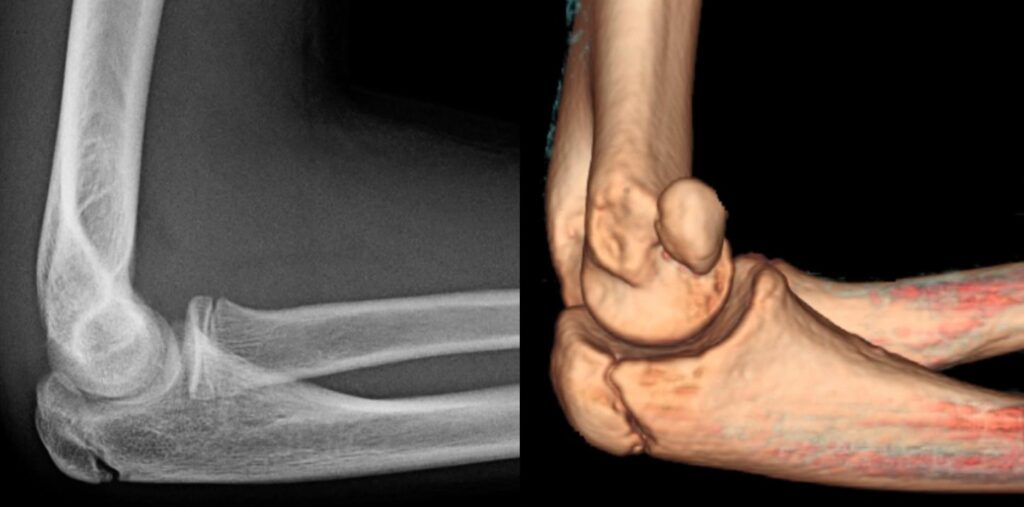
Diagnosis with Radiography
The internal epicondyle fracture is visible in a simple radiograph. If the fracture is accompanied by dislocation, the broken fragment can be seen inside the joint.
Treatment of Internal Epicondyle Fracture of the Elbow
Treatment for Non-displaced or Minimally Displaced Fractures
If the fracture is non-displaced or minimally displaced, the usual treatment involves immobilizing the elbow using a long arm splint. In this case, a minor displacement of the fractured piece is acceptable and does not require realignment. The elbow is typically placed in a splint for three weeks.
Rehabilitation After Treatment
After three weeks, the elbow joint may become stiff and have limited mobility. The patient should be under the supervision of a physiotherapy specialist and perform movements and exercises to regain the joint’s range of motion.
Treatment of Complex Fractures
If the fractured piece is trapped in the joint, the doctor will attempt to remove it using specific maneuvers without the need for surgery. If the fractured piece cannot be removed with closed maneuvers, or if the fracture is significantly displaced, surgical intervention is necessary. In this procedure, the fractured piece is fixed in its original place with pins or screws. The pins are usually removed after three weeks.
Complications of Internal Epicondyle Fractures of the Elbow
Ulnar Nerve Palsy The most significant complication of an internal epicondyle fracture of the elbow is ulnar nerve palsy. This nerve is located behind the internal epicondyle and in close proximity to it, and it may become compressed or stretched during the fracture, leading to paralysis. Fortunately, most nerve damage resulting from this fracture is temporary and usually recovers within a few weeks to months.
Need for Surgical Intervention
If there is no improvement, surgical intervention may be necessary. Sometimes, due to improper healing of the fracture and misalignment of the adjacent bones, the ulnar nerve may become compressed, causing symptoms of nerve irritation. In such cases, patients typically require surgery.
Surgical Procedure
During surgery, the passage of the ulnar nerve, which runs behind the internal epicondyle, is moved to the front to reduce pressure on the nerve and improve its function.
To make an appointment or get an online consultation with Dr. Nader Motallebi Zadeh, Limb lengthening surgeon, proceed here.