Muscle and muscle tissue is a vital component of the body’s movement system. The primary function of muscles is to generate force and create movement in the body. This movement can occur in internal organs such as the heart and digestive system, as well as in bones and limbs.
The contraction of a muscle and the application of the force it produces leads to the movement of bones. This movement can result in the overall movement of the body or a change in the position of limbs. In some cases, muscle contraction only leads to a change in muscle shape and a slight displacement, such as the contraction of the tongue muscle or the heart muscle.
Approximately 40% of the total human body weight is made up of muscles.
Types of Muscles
Based on their characteristics, muscles or muscle tissues are divided into three categories: skeletal muscles, cardiac muscles, and smooth muscles.
The contraction of skeletal muscles is voluntary and internal, while the contraction of cardiac muscle and smooth muscles is involuntary. Muscles obtain the energy they need from the oxidation of fat and carbohydrates.
Skeletal muscles are of two types:
- Type One or Slow Muscle: This type of muscle has numerous blood vessels, mitochondria, and a high amount of myoglobin, which causes the muscle to appear red. This muscle type can contract for a long duration and, in other words, has high endurance.
- Type Two or Fast Muscle: This muscle type has a high speed of contraction. It also has significant strength but tires quickly. Fast muscles can increase in volume and are usually composed of less myoglobin and mitochondria, hence they are also called white muscles.
The volumetric weight of muscles and muscle tissues is more than the weight of water, while the volumetric weight of fat tissue is less than the weight of water. Therefore, overweight individuals with more fat float better in water than those with a muscular body.
Anatomy of Muscles and Muscle Tissues
There are about 650 skeletal muscles in the human body. Muscle is part of the human body’s movement system, also known as the musculoskeletal system.
This system includes muscles, bones, joints, tendons, ligaments, and many other components. Body muscles come in various shapes and their strength varies depending on the task they perform.
The largest part of a muscle is called the origin, and the other end, where it attaches, is called the insertion. The head of the muscle closer to the body’s central axis is the origin.
Skeletal muscles in the body come in various shapes and usually end in tendons at both their ends. These tendons attach to bones and transfer the force from muscle contraction to the bones. Tendons can be round and rod-like, band-shaped, or wide and curtain-like. Around the bones, there is a layer called periosteum to which tendons are attached.
It is assumed that the origin of a muscle is attached to the bone via a tendon and does not move. With muscle contraction, the other end, i.e., the tendon attachment on the other side, moves, causing movement in the bone farther from the body’s center.
For example, the brachialis muscle in the front of the arm firmly attaches itself to the arm bone and, through its contraction, causes movement in the forearm bones.
In some cases, several muscles may have a common origin that attaches to a point on the bone and have multiple ends that attach to different points. For instance, most of the muscles in the back of the forearm attach to the lateral epicondyle of the arm at the elbow area and lower down in the forearm; each muscle attaches itself to a different point and performs a separate function.
Sometimes a muscle has one upper end and several lower ends. For example, the extensor muscles or finger extenders of the hand have one end that attaches to the lateral epicondyle of the arm bone and lowers down several tendons, each attaching to a finger. When this muscle contracts, all fingers of the hand open simultaneously.
Usually, one end of a muscle attaches to one bone, and the other end to another bone. There is typically a joint between these two bones. For example, one end of the brachialis muscle attaches to the front of the arm bone, and the other end to the ulna or lower arm bone. When this muscle contracts, the elbow joint bends. Sometimes a muscle or its tendon passes over two joints, causing movement in both.
For example, the rectus femoris muscle, located at the front of the thigh, attaches at the top to the pelvic bone and below to the tibia or shinbone. Therefore, this muscle passes over both the hip and knee joints. The contraction of the rectus femoris muscle can cause the hip joint to bend while simultaneously opening the knee joint.
Usually, several muscles are located side by side and form a muscle group or a compartment. For example, in the back of the calf, several muscles come together to form the posterior compartment of the calf. A thin but strong tissue layer called fascia covers this muscle group. This fascia is also present between the muscles in a group, allowing the muscles to move better alongside each other. Blood vessels and nerves are located next to the fascia and extend to the muscles.
Function of Muscles
The function and biomechanics of a muscle refer to the action and movement of skeletal muscles that are attached to bones. Essentially, they form a unified or integrated movement system. This system essentially functions as a lever and follows the laws of physics. As we know, every lever consists of three parts:
- The Effort Force: In the body, this force is acted by the muscle. The distance from the effort force to the fulcrum is called the effort arm.
- The Fulcrum: In the human body, the fulcrum is the joint.
- The Resistance Force: This force must be overcome to move the effort force. The distance from the resistance force to the fulcrum is called the resistance arm.
A “joint, muscle, bone” unit in most cases forms a third-class lever. In these levers, the effort force is located between the fulcrum and the resistance force, where the joint acts as the fulcrum, the bone itself is the lever, and the muscle is the effort force.
In the limbs of the body, the muscle’s attachment to the bone is usually closer to the fulcrum or joint than to the point of resistance force. For this reason, the levers in the limbs generally reduce the amount of force and increase the range of force application.
An example of this type of lever in the body is the connection of the biceps muscle to the forearm bone. Since the attachment point is closer to the elbow joint than to the palm of the hand, a short contraction of the biceps brachii muscle causes the palm to move a significant amount, but in turn, produces less force.
In some areas, such as the connection of the calf muscle to the heel bone, second-class levers are formed, where the fulcrum or joint is located between the effort force and the resistance force.
With muscle contraction, the force not only provides movement to the bone but also exerts a significant force on the joint. These forces impact the joint surfaces and may cause joint wear.
For example, during walking, significant forces impact the joint surfaces in the hip joint, so much so that these forces are often several times the weight of the individual.
When the joint surface is deformed due to factors such as fractures and is not smooth and even, the concentration of forces on the joint will be greater in some areas than others, which may accelerate joint wear.
Histology of Muscle
The histology of muscle refers to the presence of a layer called epimysium, made of connective tissue, around each muscle. This epimysium connects the muscle to the tendon and prevents friction of the muscle during contraction on another muscle or adjacent bone, and also facilitates the movements of muscles over each other.
Within the epimysium, there are several bundles called fascicles, each surrounded by a sheath called perimysium. Inside each fascicle, there are several muscle fibers.
Between the fascicles, blood vessels, and nerves are located. Each muscle fiber is a muscle cell, referred to as a myocyte. Around each fiber or muscle cell, there is a sheath called endomysium.
These sheaths, which are around the bundles of muscle cells, fascicles, and the entire muscle, provide resistance and strength to the muscle against excessive stretching and also facilitate the transfer of the force generated by muscle contraction to the tendon. These sheaths are really a kind of scaffold for muscle tissue that gives it shape and strength.
Inside each muscle cell, there are strands called myofibrils, which are actually bundles of filaments or protein threads. Myofibrils are composed of repeating units called sarcomeres along their length.
The presence of regularly repeating sarcomeres creates a striated appearance in skeletal muscles and cardiac muscles. Therefore, skeletal muscles and cardiac muscles are also called striated muscles. The filaments of the myofibrils are made of proteins called actin and myosin.
Muscle contraction is actually due to the movement and interlocking of these two proteins, actin, and myosin, alongside each other. The initiation of this contraction is triggered by a nerve signal. The nerve’s command to the muscle to contract is essentially the stimulation of the muscle by an electrical current sent from the brain through the nerve to the muscle.
This electrical stimulation initiates chemical reactions that result in the movement of the actin and myosin proteins next to each other, and with the movement of these proteins, the length of the myofibrils shortens, resulting in muscle contraction.
Muscle Strength Examination
Muscle strength examination involves assessing the capability of each muscle against a standard level of strength. If a muscle’s ability is less than the standard condition, it is measured relative to this standard.
Grades of Muscle Contraction Strength
Muscles may weaken due to various causes and diseases. To determine the extent of muscle weakness, doctors use specific criteria and divide the strength of each muscle into different grades based on these criteria.
The contraction strength of each muscle is divided into five grades:
- Grade Zero: The muscle has no contraction ability; in other words, it is paralyzed.
- Grade One: Muscle contraction can be felt, but this contraction does not cause movement in the bone or limb.
- Grade Two: Muscle contraction is strong enough to move the limb horizontally but is not sufficient to overcome the force of gravity and pull the limb upwards against gravity.
- Grade Three: Muscle contraction is strong enough to move the limb against the direction of gravity.
- Grade Four: Muscle contraction is greater than grade three but less than normal.
- Grade Five: Muscle contraction strength is normal.
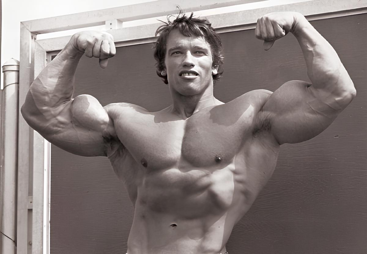
The strongest muscle in the body is the quadriceps femoris or quadriceps muscle. However, some believe that the strongest muscle is located in the buttocks area, named the gluteus maximus.
The increase in muscle size is called hypertrophy. This increase is due to the proliferation of protein filaments within the muscle cells, not due to an increase in the number of muscle cells. Muscle volume increases during puberty, and exercise can also help increase muscle size.
Reduction in size, thinning, and weakness of a muscle is referred to as atrophy. Continuous inactivity, poor nutrition, certain chronic diseases, or cancer can lead to muscle atrophy. Additionally, aging can also cause muscle atrophy.

To make an appointment or get an online consultation with Dr. Nader Motallebi Zadeh, Limb lengthening surgeon, proceed here.