In this article, we will talk about Supracondylar Elbow Fractures in Adults. Supracondylar fractures of the elbow joint in adults constitute approximately two percent of all adult fractures. This area is precisely located above the elbow joint.
- Distinctive Features of Supracondylar Elbow Fractures in Adults
- Anatomy of the Supracondylar Region of the Elbow
- Causes and Mechanisms of Supracondylar Elbow Fracture
- Symptoms of Supracondylar Elbow Fracture
- Medical Examination
- Vascular and Nerve Examination
- Diagnosis of the Fracture
- Non-Surgical Treatment of Supracondylar Elbow Fracture
- Surgical Treatment of Supracondylar Elbow Fracture
- Rehabilitation and Physiotherapy After Non-Surgical Treatment of Elbow Fracture
- Rehabilitation After Surgical Treatment of Elbow Fracture
- Types of Rehabilitation Movements
- Limitations During the Rehabilitation Period
- Outcomes of Treatment of Supracondylar Elbow Fracture
- Potential Complications of Fracture Treatment
Distinctive Features of Supracondylar Elbow Fractures in Adults
These fractures are rare in adults but are more common in children. The significance of these fractures lies in the potential damage to the brachial artery, which can be very dangerous.
Anatomy of the Supracondylar Region of the Elbow
The elbow joint is comprised of three bones: the humerus (upper arm bone) above, and two forearm bones (the ulna and the radius) below. The ulna, or lower arm bone, is located on the side of the little finger, and the radius, or upper arm bone, is on the thumb side. The ligaments of the elbow joint are located on both sides, contributing to its stability.
Causes and Mechanisms of Supracondylar Elbow Fracture
There are two main mechanisms involved in causing these fractures:
- Direct Impact: This type of fracture usually occurs when a person falls to the ground with a bent elbow, and the elbow directly hits the ground.
- Indirect Impact: This kind of fracture happens when the elbow is fully extended, and the person falls on their palm. In this case, the force is transmitted through the palm and forearm to the elbow area, causing a fracture.
Symptoms of Supracondylar Elbow Fracture
A supracondylar fracture of the elbow is primarily associated with pain. The patient usually cannot move their elbow. Other symptoms of this fracture include swelling in the elbow area, bruising, a feeling of joint instability, and in rare cases, the sensation of the sharp edge of the broken bone under the skin.
Medical Examination
To diagnose the fracture, a physician first examines the limb to check for signs like deformity, swelling, bruising, and wounds. The doctor also asks the patient to move the joints of the limb to determine if joint movement is painful. Palpating the fracture site is also an important part of the examination.
Vascular and Nerve Examination
The most crucial part of the examination is assessing the vascular and nerve status of the elbow area. A fracture in this area can damage the blood vessels feeding the forearm and hand, as well as the nerves passing through the elbow. The doctor carefully examines the function of these vessels and nerves.
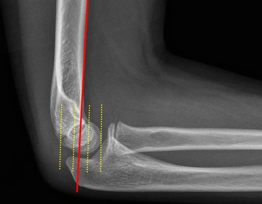
Diagnosis of the Fracture
For a definitive diagnosis of a supracondylar elbow fracture, simple radiography is used. Sometimes, an orthopedic surgeon may use a CT scan for a more detailed examination of the fracture site and to plan treatment.
The initial step in treating a fracture involves immobilizing the patient’s entire upper limb in a long splint to prevent further movement of the broken pieces. The use of localized cooling and elevating the upper limb helps to prevent increased swelling. Treatment can be either non-surgical or surgical.

To make an appointment or get an online consultation with Dr. Nader Motallebi Zadeh, Limb lengthening surgeon, proceed here.
Non-Surgical Treatment of Supracondylar Elbow Fracture
In cases where the supracondylar elbow fracture is non-displaced, the treatment primarily involves using a splint. The treating physician regularly takes X-rays of the patient’s elbow every week to ensure that the fracture does not become displaced.
If the fracture remains non-displaced for several weeks after starting treatment, the physician encourages the patient to begin moving the elbow joint. However, the patient is not allowed to lift objects for an extended period. This prolonged immobility can cause joint stiffness, and the patient needs to perform specific exercises to restore the joint’s range of motion. If the broken pieces are displaced at the start of treatment, the patient must undergo surgery.
Surgical Treatment of Supracondylar Elbow Fracture
If the broken pieces are displaced, treatment is surgical. The patient undergoes general anesthesia or anesthesia of the entire upper limb. The orthopedic surgeon accesses the fracture site by opening the skin and moving the muscles aside, then connects the broken pieces with wires, screws, or plates.
In some cases, the surgeon may relocate the nerve passing behind the elbow or break a part of the healthy elbow bone for better access to the fracture site and then reattach it with screws or pins.
In situations where the fracture is very fine and the patient is older, the doctor may decide to remove all the broken pieces and replace the elbow joint with an artificial joint.
Rehabilitation and Physiotherapy After Non-Surgical Treatment of Elbow Fracture
In cases of non-surgical treatment, rehabilitation typically begins a few weeks after the start of fracture treatment. At this stage, exercises are performed to increase the range of motion of the elbow and then to strengthen the muscles.
Rehabilitation After Surgical Treatment of Elbow Fracture
If the fracture has been treated surgically, the movement of the elbow joint should start much earlier to prevent joint stiffness. Usually, elbow movements start the day after surgery, and the patient should perform these movements every day under medical supervision.
Types of Rehabilitation Movements
Initially, active movements are performed, where the patient bends and straightens the elbow using their own muscular strength. After some time, passive movements are added to the rehabilitation, where the patient uses the other hand or another person to stretch the injured limb to open the elbow.
In later stages, exercises to strengthen the muscles of the arm and forearm are added to the rehabilitation program.
Limitations During the Rehabilitation Period
The patient should not lift heavy objects with the injured hand for 6 to 12 weeks. If an elbow joint replacement has been performed, the patient must adhere to specific limitations set by the doctor for the rest of their life regarding lifting heavy objects.
Outcomes of Treatment of Supracondylar Elbow Fracture
The goal of treating supracondylar elbow fractures is to heal the fracture in an optimal position so the patient can restore the elbow joint’s movements to its pre-injury state. Full recovery usually takes one to two years, but patients can generally return to their previous activities after six months.
To make an appointment or get an online consultation with Dr. Nader Motallebi Zadeh, Limb lengthening surgeon, proceed here.
Potential Complications of Fracture Treatment
- Surgical site infection: This complication is rare but problematic, especially if the infection involves the bone.
- Nerve damage: After a fracture, one of the elbow’s nerves may be affected, which usually improves after a few weeks.
- Non-union of fracture: Sometimes, despite surgery, the fracture does not heal. Possible reasons include not following medical instructions, underlying diseases like diabetes, or smoking.
- Limited elbow movement: Some patients may not be able to fully bend or straighten their elbow. The extent of limitation depends on various factors.
- Production of extra bone: Sometimes extra bones form around the elbow, which can cause movement limitations.
- Arthritis due to fracture: Osteoarthritis is also a complication of supracondylar elbow fractures. This condition can cause pain and limited elbow movement, though it does not always lead to daily activity problems.